
Research
We are working on solutions to a number of diverse problems.Dyssynchrony in Patients with Heart Failure and Congenital Heart Disease
Project Details
Normal hearts contract in synchrony to optimize function - all the muscle in the ventricles typically contract within a period of about 50 milliseconds. However, approximately 30% of patients with heart failure have a conduction block in the heart which leads to dyssynchronous contraction. Dyssynchrony can be diagnosed using many different imaging modalities including echocardiography and cardiac magnetic resonance imaging. Many of the projects in our lab focus on the diagnosis of dyssynchrony in the heart. We are particularly interested in the role of dyssynchrony in patients with congenital heart disease.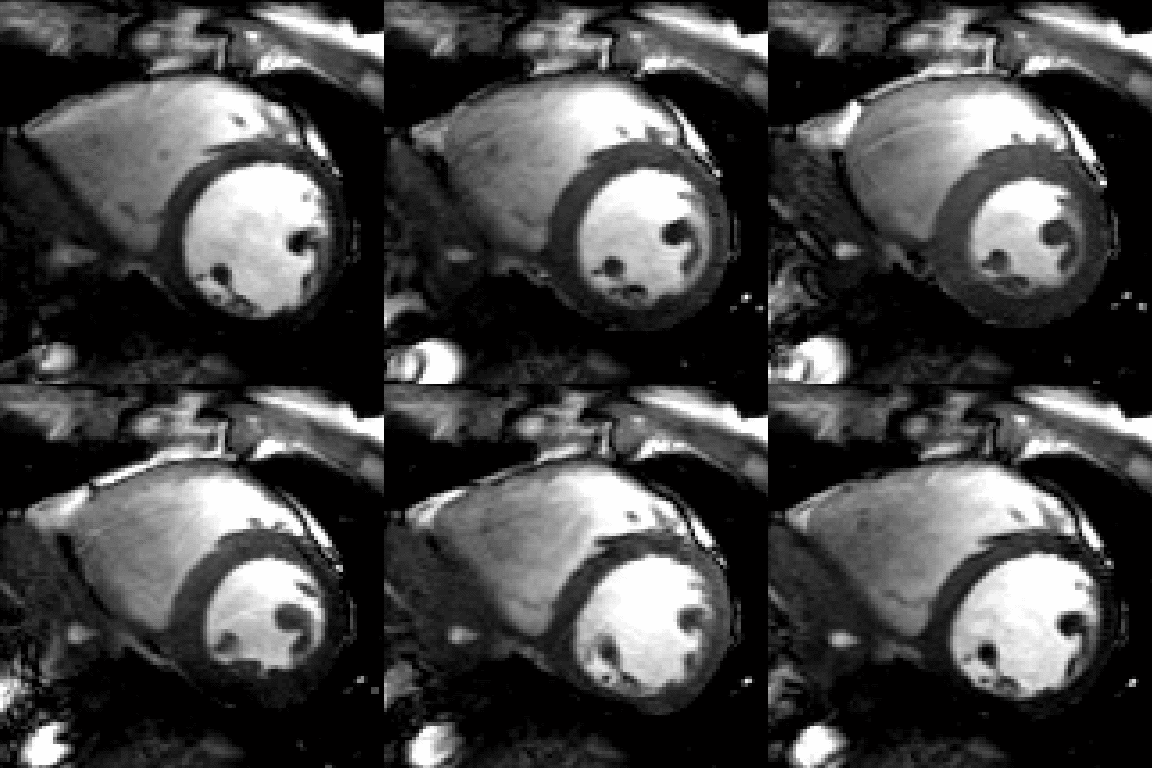
Short-axis cardiac magnetic resonance image (MRI) of a normal, healthy subject
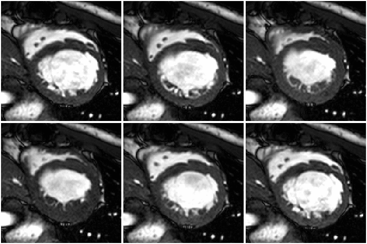
Short-axis cardiac magnetic resonance image (MRI) of a child with heart failure and dyssynchrony
3-Dimensional Modeling of the Heart from Cardiac MRI
Project Details
Our lab uses cardiac magnetic resonance imaging (MRI) to reconstruct 3-dimensional models of the heart. We use these models to help us understand cardiac function in both normal subjects and patients with heart disease (such as congenital heart disease or heart failure).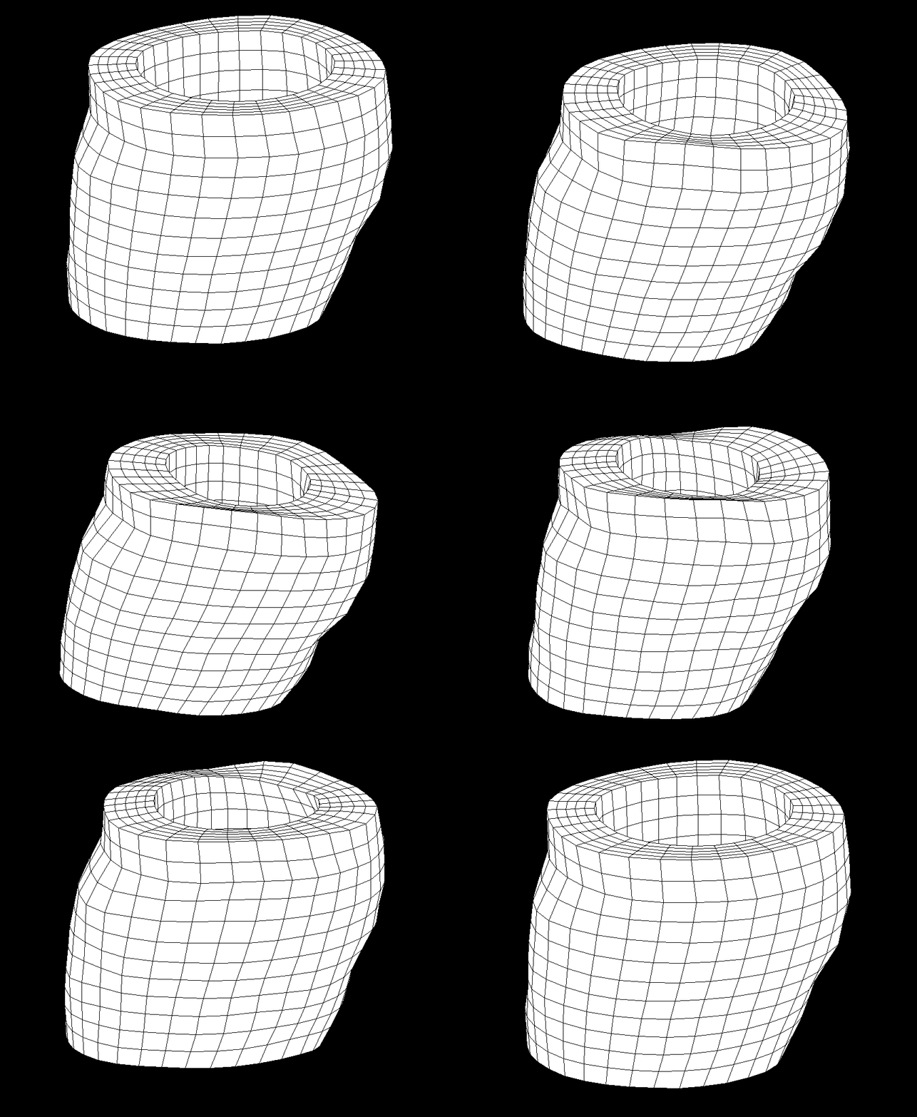
3-Dimensional model of the left ventricle
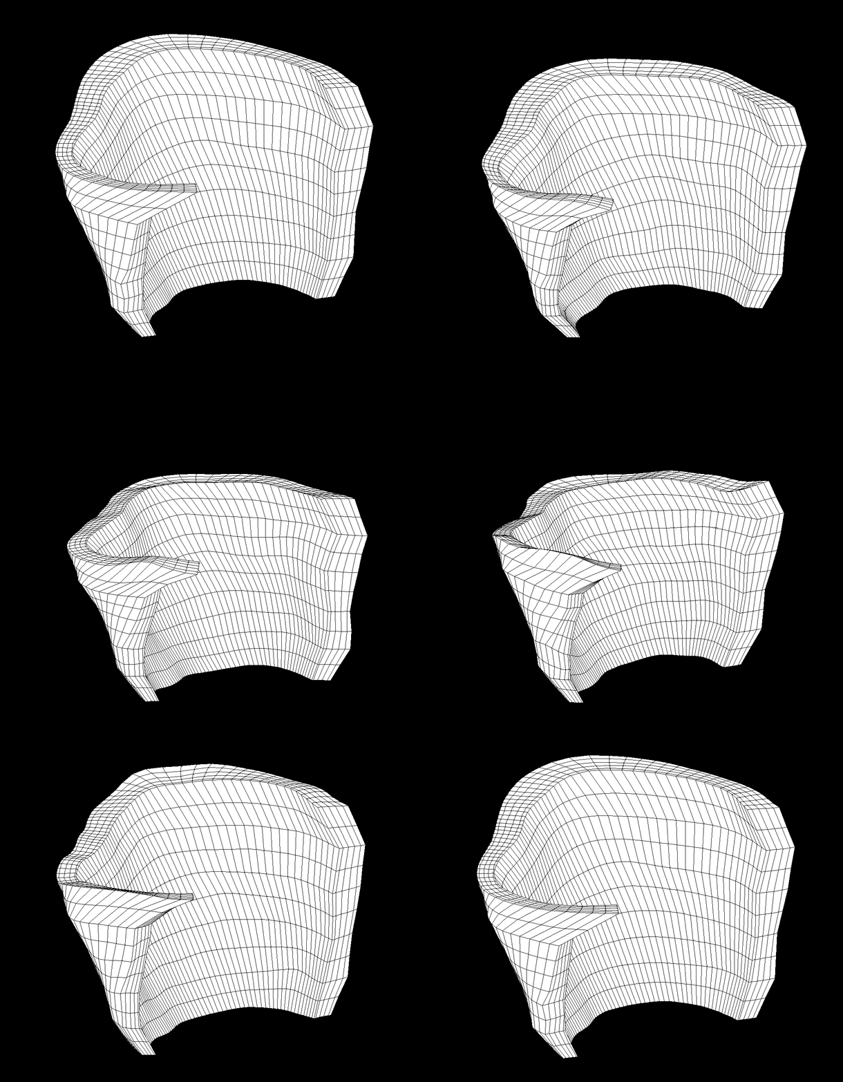
3-Dimensional model of the right ventricle
Cardiac Motion and Mechanics of the Human Heart Using DENSE
Project Details
We use Displacement Encoding with Stimulated Echoes (DENSE), a specific cardiac magnetic resonance imaging protocol, in humans to help us better understand the patterns of cardiac motion in healthy subjects and patients with heart disease.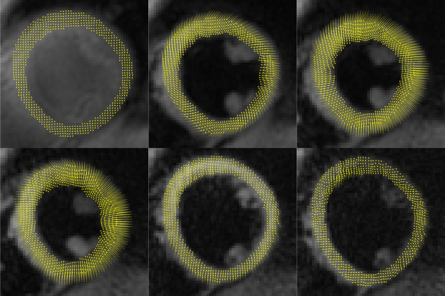
Motion trajectories of the left ventricle
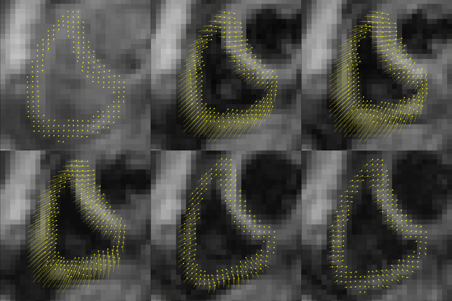
Motion trajectories of the right ventricle
Cardiac Motion and Mechanics of the Human Heart Using Tagged MRI
Project Details
We use a technique called CSPAMM (or "tags") to help us better understand the patterns of cardiac motion in healthy subjects and patients with heart disease.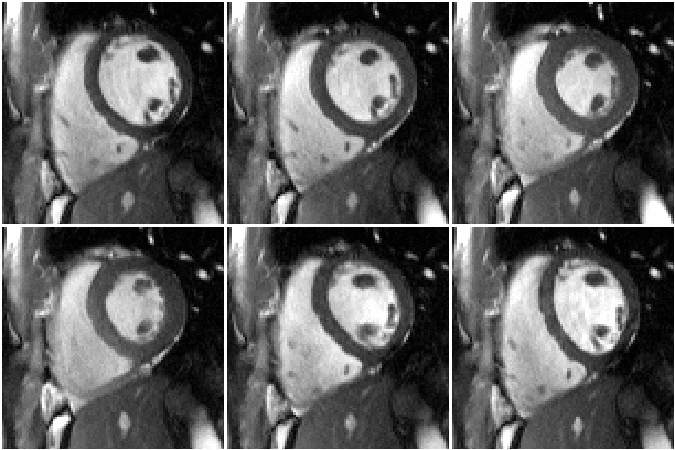
Standard short-axis image without tags
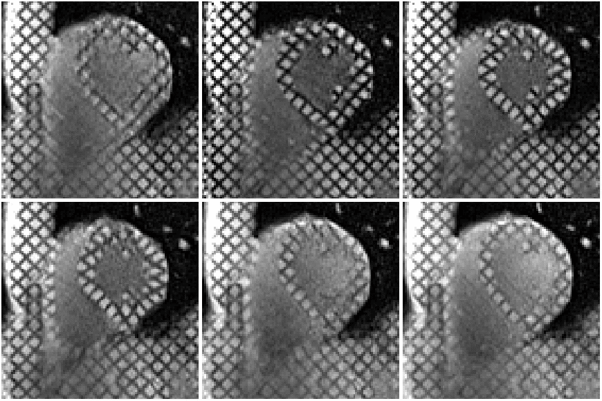
Short-axis image in the same location using tags to look at cardiac motion and deformation
High Resolution DENSE to Improve Quantification of Cardiac Motion
Project Details
We use Displacement Encoding with Stimulated Echoes (DENSE), a specific cardiac magnetic resonance imaging protocol, in humans to help us better understand the patterns of cardiac motion in healthy subjects and patients with heart disease.
Standard resolution DENSE MRI of the heart

High resolution DENSE MRI of the heart
3D Reconstructions of Aortic Aneurysms in Mouse Models
Project Details
We use cardiac magnetic resonance imaging (MRI) to reconstruct 3-dimensional models of the aorta in mouse models. We use these models to help us better understand the mechanisms and potential therapies for human aortic aneurysms.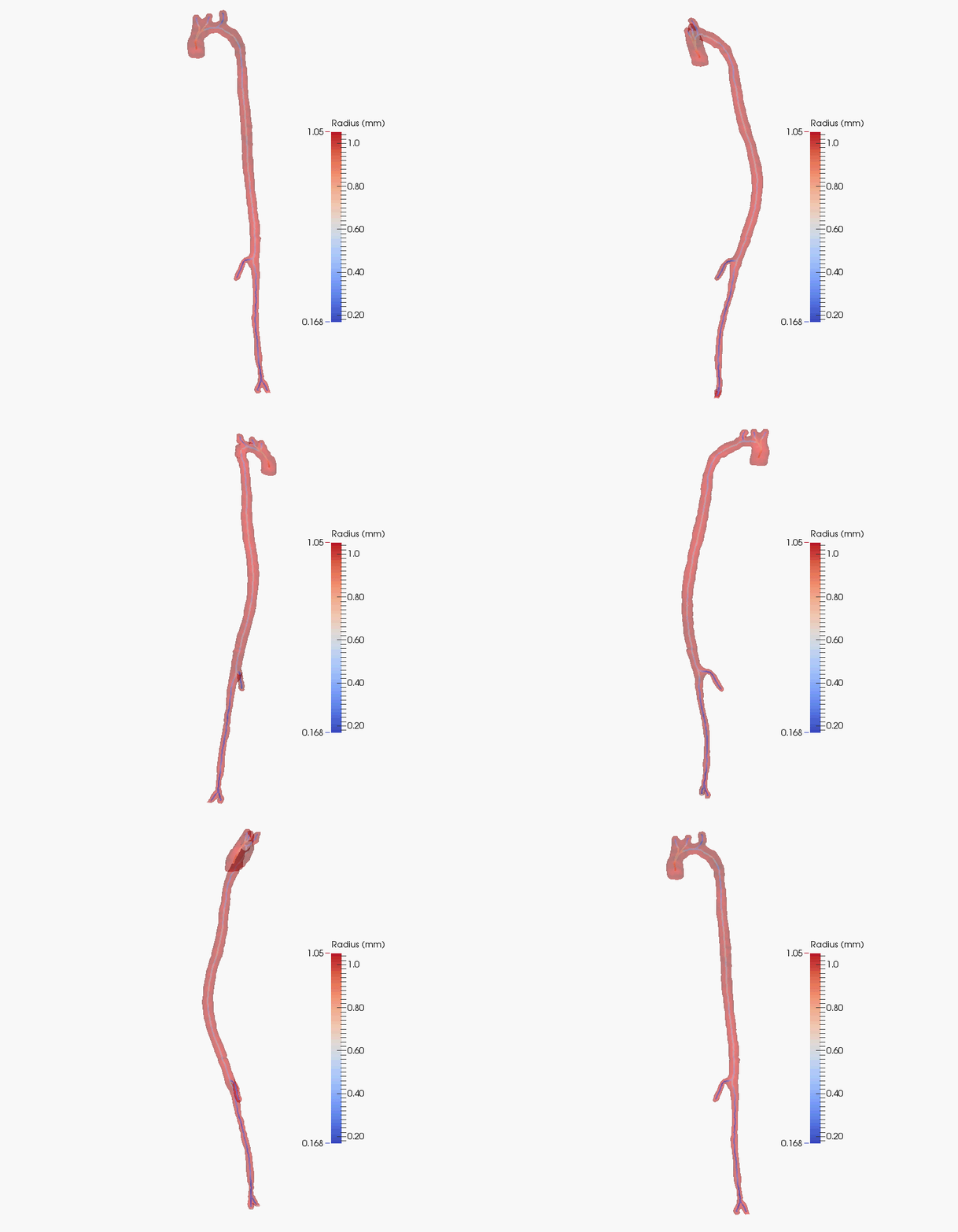
3-Dimensional reconstruction of a normal mouse aorta
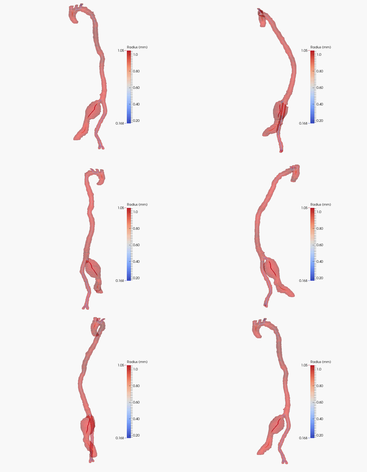
3-Dimensional reconstruction of the aorta from a mouse with a genetic alteration which causes an aneursym
Cardiac MRI in Mouse Models
Project Details
We use cardiac magnetic resonance imaging (MRI) in mouse models to help us better understand the mechanisms and potential therapies for human heart disease.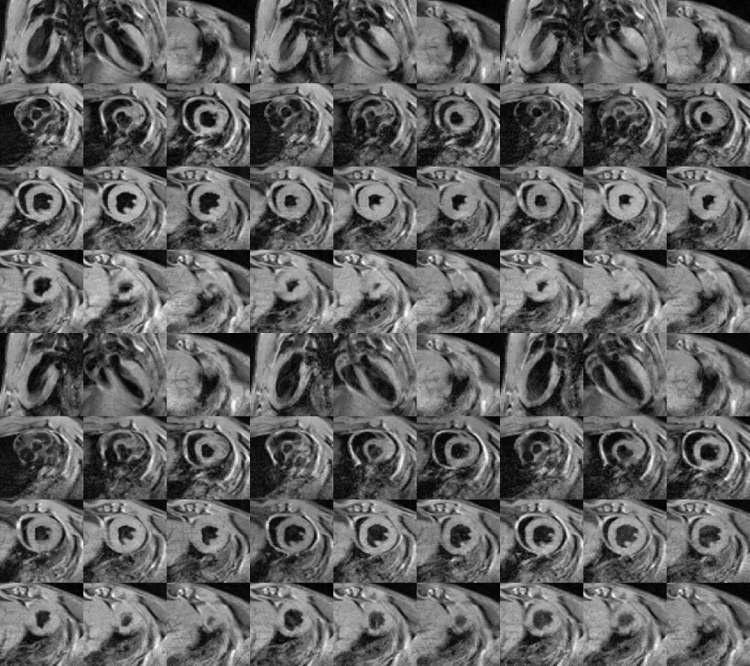
Cardiac MRI of a mouse heart
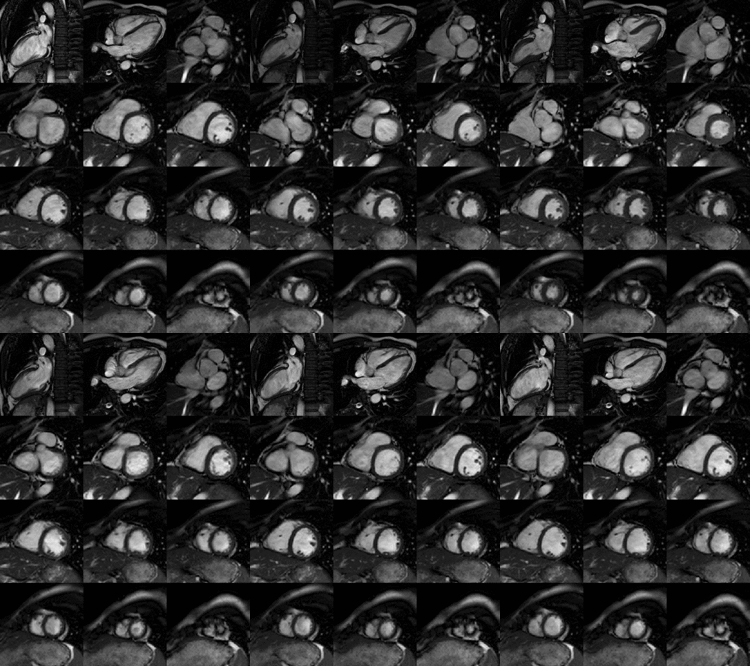
Cardiac MRI of a human heart
Cardiac Imaging Research Lab
Center for Health Research
Geisinger Clinic
100 North Academy Avenue
Danville, PA 17822-4400
Email: bkfornwalt@geisinger.edu
Center for Health Research
Geisinger Clinic
100 North Academy Avenue
Danville, PA 17822-4400
Email: bkfornwalt@geisinger.edu